Three decades supporting the flow of health and medical research discovery
18 May 2026
On most days at SAHMRI, the seeds of future breakthroughs are being quietly planted. Somewhere inside each sample could be the answers to shape a thesis, a clinical trial or even a new treatment, but before any of that can come to fruition the right cells need to be identified and interpreted.
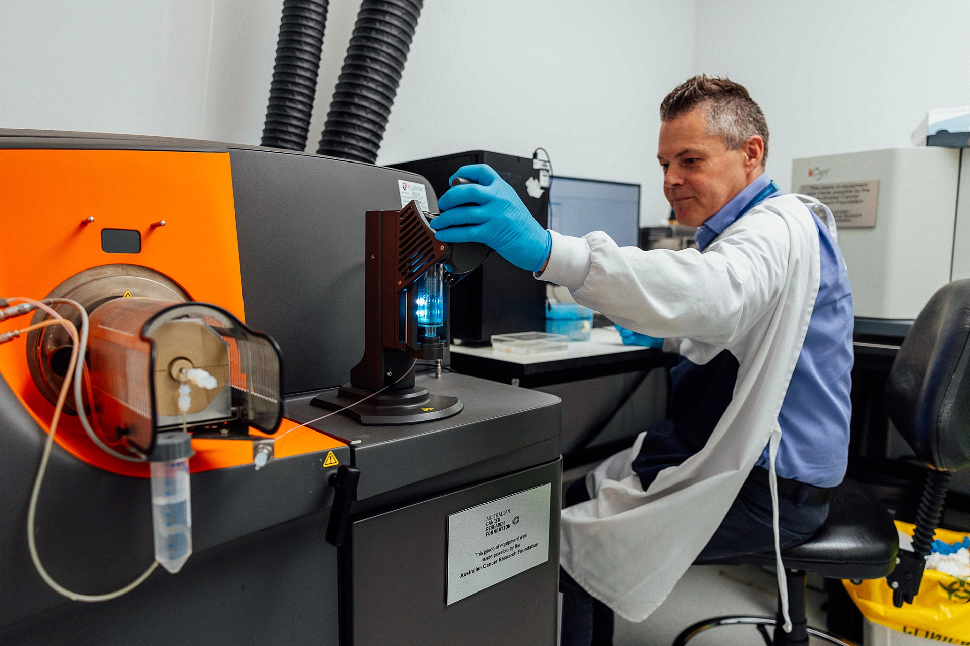
That moment when complexity gets refined to clarity is where Dr Randall Grose has built his career. For more than three decades, he’s worked in the engine room of health and medical research, using flow cytometry to turn biological noise into insight and helping generations of researchers see what was previously invisible.
“I’ve been at this since the late 1900s,” Dr Grose says with a wry grin, a tongue-in-cheek nod to the fact he’s been working at the coal face of flow cytometry for what’s felt like an age.
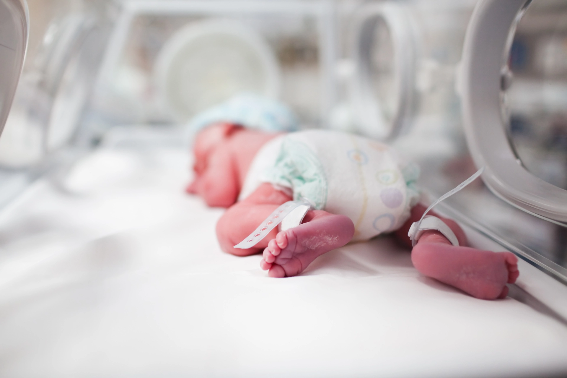
“I did my Honours at the Women’s and Children’s Hospital in the 1990s,” he says. “Back in those days people were asking if there’d ever be a real use for these machines. The thinking was maybe each state would have one cytometer. Now there are dozens just in South Australia.”
Flow cytometry is elegant, powerful and highly intricate, but Dr Grose distils all the complexity down to essentially, “passing cells one at a time through a laser beam and measuring the light they scatter and emit, to reveal what kind of cell it is and what it’s doing.”
Throughout his career, the raw concept hasn’t changed, but the tech has evolved out of sight, greatly increasing accessibility and efficiency.
“What we do now compared to 25 years ago is like going from black and white TV to high definition. Back then you might have had one or two lasers and a handful of markers. Now we can collect an enormous amount of information from every single cell.”
Modern flow cytometry is not just about detecting and measuring the cells, however. Those cells can also be automatically sorted by type or characteristics and saved for future investigation. The basic idea behind sorting cells is older than most people think.
“It actually comes from inkjet printers—you charge a stream of droplets and deflect them between plates. That same physics became a way to separate living cells.”
Flow cytometry has been at the heart of Dr Grose’s research since his PhD. After his doctorate, he stayed in Adelaide and kept pushing the method into more practical territory.
“A lot of the work was about taking very rare cells and understanding how they behave or isolating them so they could be used in the next step,” he says. “To do that, the cells have to stay alive, which makes the sorting process critical.”
In the 2010s Dr Grose’s career took him to Chicago, where he gained valuable experience working in a hospital linked, clinical manufacturing environment.
“It showed what’s involved when you start thinking about scale, timing and real world constraints. It reinforced how important careful, high quality sorting is when you’re working with limited material.”
He returned to Adelaide in 2014 to lead SAHMRI’s flow cytometry capability as manager of the Australian Cancer Research Foundation (ACRF) Cellular Imaging and Cytometry Core Facility. “When I started, we had two analysers and one cell sorter,” he says. “Over the years we’ve grown steadily, added more instruments and significantly expanded the research we can support.”
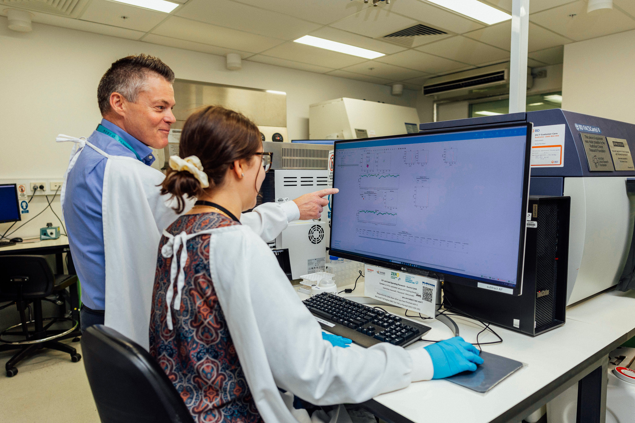
Today, the facility performs crucial analysis for hundreds of clients across SAHMRI and the wider biomedical precinct including the universities, major hospitals and industry.
“Some researchers just want a simple answer, is something there or not? Others are asking much more complex questions and collecting a lot of information from each cell.”
“Samples are extremely valuable, and researchers typically aim to extract as much information as possible from them, especially since some samples can only be collected once,” Dr Grose says. “They hand us their samples, we do the sorting or analysis, and we give them back the cells and the data so they can move forward with their research.”
A common scenario involves experiments where only a small fraction of cells behave the way researchers want. “You rarely get a perfect result straight away. Often only a small percentage of cells are useful. If you don’t separate them, the experiment can’t progress. Sorting lets us give researchers a clean starting point.”
One of the biggest steps forward has been moving into newer instruments that capture far more signal from each sample. “Older systems looked at narrow slices of light, newer ones capture everything that’s coming off the cell,” he says. “That means you can combine reagents you couldn’t before and learn much more from the same sample.”
Philanthropic support has been crucial to making that progress possible. “There’s a long history of support for flow cytometry in South Australia,” he says. “Over the years, supporters including the ACRF, Detmold Group and McMahon Services have helped drive significant investment into research and development, training and fellowships. Without that support, we might not still be here.”
“It allows us to acquire new technologies, support groups that don’t have their own labs and take on work we simply couldn’t do otherwise.”
“The equipment we use is not only costly to acquire but also expensive to maintain. Facilities like ours play a crucial role in making these machines accessible to all researchers.”
Like many core facilities, flow cytometry at SAHMRI runs on a cost recovery model that heavily subsidises internal users. “If we charged the true cost, it’d deter people and slow down a lot of research,” he says. “The point is to keep discovery moving. External and commercial users pay full price, but internally we work hard to keep access affordable.”
For Dr Grose, the most satisfying moments are the subtle ones. “When someone stops you in the lab and says their cells recovered and their experiment worked, that’s great,” he says. “You hear about projects long before they’re polished and published. You see the science taking shape.”
Looking ahead, he’s focused on keeping pace with the ever-evolving landscape. “The next generation of cytometers will let you actually image the cells as well as measure them. You don’t want to get left behind,” he says. “The benefits of funding flow cytometry at any time are long lasting, but you’ve got to continually invest to stay at the forefront.”
Appropriately named, flow cytometry truly does flow through the stages of research at SAHMRI, from inception to clinical translation, all powered by philanthropic giving.
“Funding flow cytometry funds possibility. It turns a precious sample into answers, a good idea into data, and a stalled project into momentum.”
Through countless hours of quiet, meticulous effort, Dr Randall Grose keeps doing his part to drive health and medical research forward from behind the scenes. He plans to continue going with the flow to see where it takes him, one cell at a time.
Contribute to research at SAHMRI
Donate todayRelated Articles


SAHMRI is located on Kaurna Country. We pay respects to the Kaurna people of the Adelaide Plains and to all Aboriginal and Torres Strait Islander people. We are committed to embracing knowledge and culture as we continue our working journey to incorporate Aboriginal health research across all of our themes and further reconciliation.
Read more"Tirki nguta tappa tangka marninithi"
(Learning and knowledge are the pathway to alter the mind)
- Uncle Lewis Yerloburka O’Brien, Kaurna Elder